
According to Routt ML Jr, Simonian PT, Agnew SG, Mann FA; Radiographic recognition of the sacral alar slope for optimal placement of iliosacral screws: a cadaveric and clinical study.; J Orthop Trauma 1996;10(3):171-7:
"Malpositioning of sacral screws happens more often when common variations in the morphology of the upper sacral segments are unrecognized. We sought to delineate readily reproducible radiographic anatomical clues of upper sacral morphology to improve iliosacral screw placement.
. . . Materials and methods of the cadaveric study: Radiological anatomical correlations of sacral anatomy were studied in 10 fresh frozen cadaveric pelves without evidence of skeletal disease, obtained from six male and four female donors.
. . . Results of a cadaveric study: Gross inspection of the upper sacral segments of three specimens found three specimens with morphologies different than the other seven. In these three:
1) the sacral alar were angulated acutely"
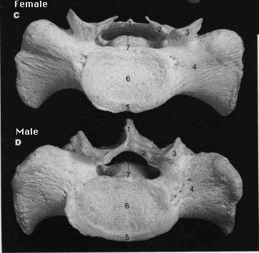
from McMinn RMH, Hutchings RT; Color Atlas of Human Anatomy, 2nd edition ; p80; Year Book Medical Publishers, Chicago 1988
The sacral alar slope appears to be typically more acutely angulated in the female.
2) the SI joints were undulating "tongue in groove" surfaces

from Gray's anatomy
Undulating "tongue in groove" surfaces seem to typify articular surfaces of the female SI joint.
3) the first sacral vertebral segment was located at the same level of the iliac crests.

from Gray's anatomy
Relative to the male pelvis, the level of the first sacral body seems to be closer to the level of the iliac crests in the female pelvis.
4) the first sacral vertebral segment is not recessed within the pelvis.

from Gray's anatomy
Relative to the male pelvis, the level of the first sacral body seems not to be recessed within the pelvis (in order to maximize the AP diameter of the birth canal in the female pelvis).
5) Mammillary proceses are noted along the alae.

from Gray's anatomy
No references can be found with respect to sacral mammillary bodies. Relative to the male pelvis, there are rounded prominences on the superior-posterior alar surface in the female pelvis.
from McMinn RMH, Hutchings RT; Color Atlas of Human Anatomy, 2nd edition ; p80; Year Book Medical Publishers, Chicago 1988
Conclusion:
Perhaps the hallmarks of "upper sacral dysplasia" are, in fact, normal variances of the female sacrum.