Date: Tue, 2 Apr 2002 23:33:40 -0000
Subject: Ankle Subluxation
From: Dr. Josep M. Muñoz Vives
I wish to present a case to the list.
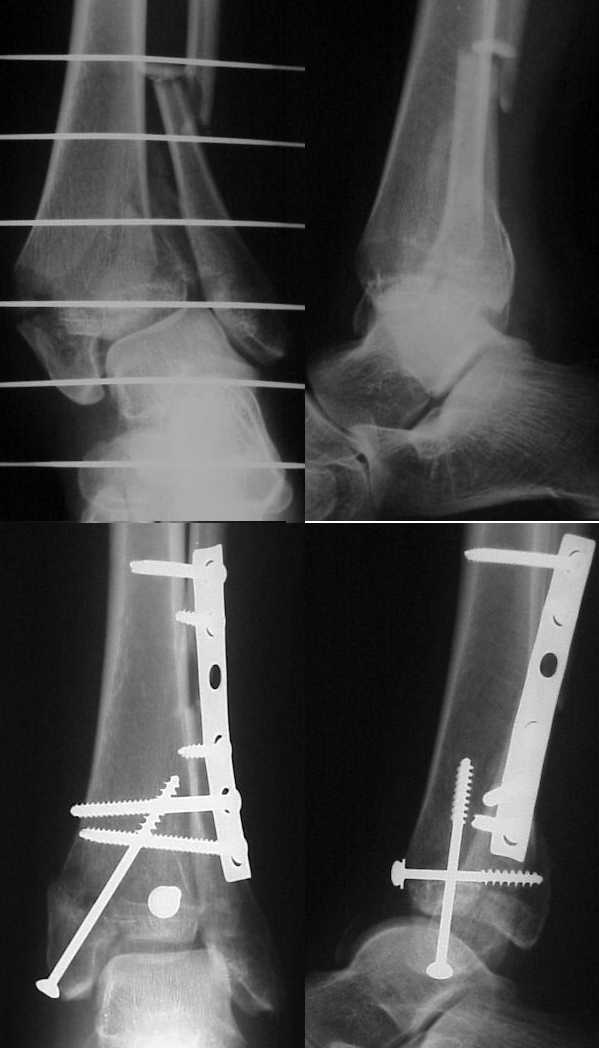
20 yo woman was injured in a MVA. The fracture was aligned in a padded cast and surgery was delayed for a week because of severe swelling. A week later, open reduction and internal fixation of the fibula with tibio-fibular screws and percutaneous synthesis of the tibial pilon was done. After surgery the leg was splinted in a soft padded cast. During surgery, fluoroscopy was used, but the subluxation was unnoticed.
The subluxation was appreciated in the post-op x-ray hardcopies. It was attributed to the cast, but the cast was released and the subluxation persisted. The patient can move her ankle in an almost normal range of motion with no pain and the subluxation is fixed in all the range. A CT of the mortise and syndesmosis shows that the fibula is in the right place. A x-ray of the other ankle is normal.
|
All coments will be welcomed.
Dr. Josep M. Muñoz Vives
Orthopedic Dept.
Hospital Universitari Dr. Josep Trueta.
Girona
Catalunya, Spain
Date: Wed, 3 Apr 2002 10:23:45 -0800
From: Carlo Bellabarba
Dr. Vivez,
This is an interesting situation. The postoperative lateral xray suggests a considerable apex posterior angulation of the fibula at the fracture site. This may be a major contributor to the problem, although I can't tell for sure on the available xrays. Do you have a lateral view that shows more of the fibula?
Carlo Bellabarba
Harborview Medical Center Seattle, WA
Date: Wed, 3 Apr 2002 21:34:13 +0200
From: Dr. Josep M. Muñoz Vives
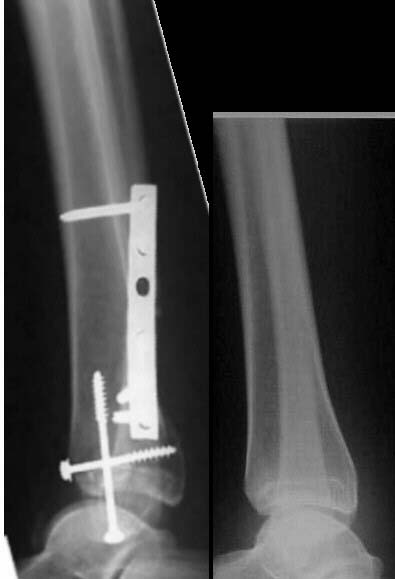 |
Relooking at the x-rays I agree with Dr. Bellabarba that there may be some angulation at fracture site, although there seems that at the syndesmosis the fibula is located were it should be comparing to the healthy side. I've already examined the patient under image intensifier (fluoroscopy) and she has a near complete unpainful range of motion, but the subluxation does not correct with dorsiflexion (even forced).
Dr. Josep M. Muñoz Vives
Orthopedic Dept.
Hospital Universitari Dr. Josep Trueta.
Girona
Catalunya, Spain
Date: Wed, 03 Apr 2002 15:25:29 -0500
From: James Carr
In looking at the x-ray sent, it does appear that the fibula does have apex posterior angulation, as was previously noted. I would wager this would have to be corrected. Someone also mentioned the medial side, but I couldn't tell based on the images. The case I alluded to with the anterior subluxation would not reduce even with open reduction. There were no bony fx, and I only obtained partial correction.
JC
Date: Wed, 3 Apr 2002 19:32:12 +0100
From: chris wilson
The medial malleolus is not reduced. It is in valgus and probably a little anterior, but it may be that the mortice is too small and is effectively extruding the talus anteriorly in neutral or plantarflexion. Screen it under fluoro and see if it drops back into position on dorsiflexion. You might have to re-do the medial fixation.
Chris Wilson
Knee and Trauma Surgeon
University Hospital
Cardiff
UK
Date: Wed, 03 Apr 2002 14:13:30 -0500
From: James Carr
Assuming the bones are in good position, there are two possibilities:
1. The anterior tib-fib ligament is torn, and the ankle was subluxated at the time of splinting- i.e. holding the big toe to put the post-op splint on.
2. The syndesmosis is overtightened, along with tear of the ATF ligament. I once had one of these that was nearly impossible to reduce 6 weeks later.
I would first try exam under anesthesia, and hopefully reduce closed with the foot maximally dorsiflexed.
James B. Carr, MD
Palmetto Health Orthopedics
Date: Wed, 03 Apr 2002 17:14:06 -0500
From: Kevin Pugh
Great case.
I think that those who have responded to date have identified the issues. The fibula appears to be angulated. Thus is may be in the right place in relation to the tibia, it is angulated, and you wouldn't see that on an axial cut. The medial malleolus is also malreduced, and effectively in valgus. The mortise does not appear to be wide enough. Do you have a true mortise view?
If the soft tissues are now adequate, you may wish to go to the OR and do a more extensive procedure to correct the issue...rereduce the medial side +/- the fibula.
If you don't get this soon, it will be very hard to do it after the soft tissue envelope contracts.
Kevin J. Pugh, MD
Chief, Division of Trauma
Department of Orthopaedics
The Ohio State University
Columbus, OH 43210
Date: Thu, 04 Apr 2002 11:18:25 -0500
From: OTS1
this is a very typical case and I actually did one of these last week and video taped the entire intraoperative findings. My patient had a similar injury and a segmental fibula. Despite orif of the ankle and reconstruction of the syndesmosis, the ankle was still grossly subluxable. This is because the entire capsule is ripped posteriorly and laterally. If you extend your lateral incision distally, you can then see that the lateral collateral ligament is completely disrupted. If you go ahead and fix that, either directly or with mitek sutures, your ankle will be completely stable!
roy sanders, m.d.
tampa, fl.
Date: Thu, 04 Apr 2002 10:29:42 -0600
From: Steven Rabin
i've just seen a similar case last week but it is at 4 months from injury. Is it worth it to try a ligament repair now, or is a more extensive reconstruction indicated?
Date: Thu, 04 Apr 2002 23:14:56 +0100
From: Peter Hamilton
Looking at the films provided it appears that malreduction has lead to a narrowed mortise and the talus has been extruded anteriorly as suggested. When this is rectified if there is still soft tissue instability a temporary calcaneo-talo-tibial pin may be useful while the tissues contract or a short term ex fix.
Good Luck.
Mr. Peter Hamilton MBBS(Hons.),FRACS(Orth).
Trauma & Orthopaedic Surgeon
Addenbookes Hospital
Hills Rd
Cambridge CB2 2QQ
United Kingdom
Date: Thu, 04 Apr 2002 18:15:01 +0000
From: b.meinhard
I have treated several patients with this constellation of injury. They have often been open at the time of injury. Correction of the angulated fibula and ligamentous reconstruction at the time of revision surgery is indicated. Furthermore one must be certain that there is no soft tissue interposition which would impede reduction at the next surgical procedure. What I do not like to see is a tibio talar pin holding reduction instead of surgical repair of the ligaments and capsule when feasible.
Date: Thu, 04 Apr 2002 18:28:18 -0600
From: Steven Rabin
i've used the temporary calcaneo-talo-tibio pin technique before and not been happy with it, which is why my previous email asked roy and others what other options they would do. if you use this kind of technique, put the pin in obliquely so it exits the tibial cortex and doesn't end up intramedullary, since it can break and if it is intramedullary it will not be retrievable easily, but if it goes through the cortex, it can be removed. (if it breaks, it will break in the ankle joint and require removal.) this is a problem that occurs more often than most people would think in these high energy ankle fracture dislocations.
Date: Sat, 6 Apr 2002 07:08:37 +0100
From: Rajesh
Looking at the postop Xrays, I think the problem with the medial malleolus has come up because it has been "reduced" to align the outer tibial cortex and not the joint surface. Normally this would not be a problem butif there is comminution of the cortex, then we would be reducing it at a higher level with respect to the articular surface. It is quite easy to check articular surface alignment through the anteromedial corner of the arthrotomy. The mortice has also been narrowed, effectively pushing the wider portion of the talus out of the mortise. If it will not reduce on dorsiflexion (while screening) I think you are better off taking down the fixation and redoingit, taking care to address the problems identified.
rajesh