Surgical Technique for ZMS Recon Nail
Incision and Exposure
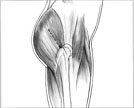 An incision is madebeginning at the tip of the greater trochanter extending proximally 10 cm(click image to enlarge). The fascia of the gluteus maximus isincised in line with its fibers. The subfascial plane of the gluteus mediusis identified, and the trochanteric fossa is palpated. The muscles are retractedto facilitate palpation or visualization of the piriformis fossa.
This may be difficult in the obese patient, especially if flexion andabduction of the proximal fragment causes the tip of the trochanter to lieagainst the ilium. Positioning techniques used to expose the tip of thetrochanter include adduction of the leg and positioning of the patient'storso away from the affected extremity.


